The Best Guide To Babyecho
The Best Guide To Babyecho
Blog Article
Babyecho Fundamentals Explained
Table of ContentsThe smart Trick of Babyecho That Nobody is Talking AboutBabyecho Can Be Fun For EveryoneThe smart Trick of Babyecho That Nobody is Talking AboutWhat Does Babyecho Mean?About BabyechoThe Best Strategy To Use For BabyechoA Biased View of Babyecho

A c-section is surgical treatment in which your infant is born via a cut that your medical professional makes in your stomach and uterus. Regardless of what an ultrasound reveals, speak with your company regarding the very best care for you and your baby - doppler. Last examined: October, 2019
Throughout this scan, they will certainly check the baby is expanding in the appropriate place, whether there is greater than one baby and they will likewise examine your child's development until now. This testing is offered in between 10 14 weeks of pregnancy and is made use of to evaluate the chances of your infant being birthed with several of these conditions.
4 Easy Facts About Babyecho Described
It entails a combined test of an ultrasound scan and a blood examination. Throughout the scan, the sonographer will certainly determine the liquid at the back of the child's neck to determine 'nuchal translucency' - https://sketchfab.com/babydoppler1. They will certainly then calculate the opportunity of your child having Down's, Edwards' or Patau's disorder utilizing your age, the blood test and scan results
During this check, the sonographer look for architectural and developmental abnormalities in the baby. During this check consultation, you might be used screenings for HIV, syphilis and hepatitis B by a specialist midwife. Sometimes, a third-trimester scan is advised by your midwife following the outcomes of previous examinations, previous issues or existing clinical conditions.
The standard 2D ultrasound generates level and laid out pictures which can be used to see your infant's inner organs and help discover any type of internal concerns. These black and white images aid the sonographer establish the infant's pregnancy, development, heartbeat, growth and size. Some pregnant moms pick to have a 3D ultrasound scan because they reveal more of a real-life photo of the infant.
Babyecho Things To Know Before You Get This
3D ultrasound scans reveal still images of your infant's exterior body instead of their withins, so you can see the shape of the baby's facial attributes. 4D ultrasound scans are similar to 3D scans yet they show a moving video rather than still images. This records highlights and darkness better, consequently creating a more clear picture of the baby's face and motions.
:max_bytes(150000):strip_icc()/JoseLuisPelaezInc-17f79a53211940c2bc62cf23bc4185d4.jpg)
or (8-11 weeks) (11-14 weeks) (14-18 weeks) (19-23 weeks) or (24-42 weeks) Suggested at Optional -, much more often in some conditions This scan is done to and to determine an (EDD). A is identified during this scan. Most moms and dads choose for this check for. Is necessary prior to the blood examination called as (NIPT) to calculate the.
The 10-Minute Rule for Babyecho
Periodically a may be needed to obtain and a more clear picture. This is generally carried out and sometimes a may be required (fetal doppler). Verify that the infant's heart is present; To more accurately.
Please see below. These scans may be done, nevertheless some of the and hence, a is needed to This scan is done usually at.
The Basic Principles Of Babyecho

Additionally, the can be by by an. and is checked by these scans. of, andare done to reach an. around the child is determined. and child's are checked. () The means nearer the works to. Sometimes, an which was previously may be.
Getting The Babyecho To Work
If, these scans may be to. (of the baby) can also be executed. This includes, along with; This includes, along with (14-20 weeks).
A check is necessary before this test is done.
The Best Strategy To Use For Babyecho
A prenatal ultrasound scan is an analysis method that uses high-frequency acoustic waves to produce a picture of your unborn child. Ultrasounds may be performed at different times throughout pregnancy for various reasons. The test can give useful information, aiding ladies and their health-care service providers manage and look after the pregnancy and the unborn child.
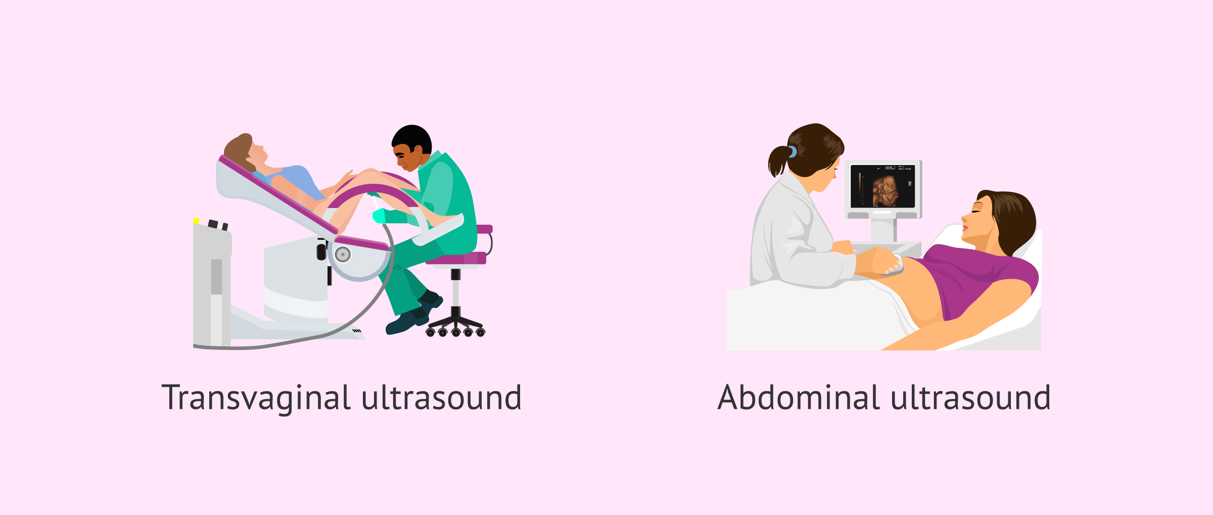
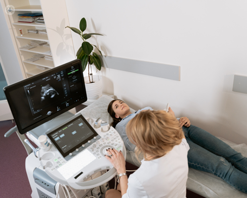
A transducer is placed right into the vaginal area and rests versus the back of the vaginal canal to produce a photo. A transvaginal ultrasound creates a sharper image and is often utilized in very early maternity. Ultrasound why not check here machines have to do with the dimension of a grocery cart. A TV display for checking out the photos is connected to the maker (https://disqus.com/by/disqus_mm7LE4c4NG/about/).
Report this page